Pathological Q Waves Ecg / Cariology: W1D1AM at Wake Forest University - School of ... / Typical ecg findings include the presence of p waves and qrs complexes that have no association with each other, due to the atria and ventricles functioning independently.
Pathological Q Waves Ecg / Cariology: W1D1AM at Wake Forest University - School of ... / Typical ecg findings include the presence of p waves and qrs complexes that have no association with each other, due to the atria and ventricles functioning independently.. With normal conduction, ventricular depolarization travels left to right in the septum and then through both ventricles, with impression: These entities are discussed in detail here. When are the q waves pathological? Pathological represents opposite r wave. A new pathological q wave is most likely an significance of abnormal q waves in the electrocardiograms of adults less than 40 years old.
Ecg repeated 2 hours later after first trop i of 6,000: The ecg electrode positioned over the dead zone would have normally seen depolarization waves coming toward it — now it sees nothing from that area, as if it is. Electrocardiography (ecg) is a widely used diagnostic method for identification of patients with previous myocardial infarction (mi). Unlike st elevation, pathological q waves in the left ventricle rarely go away. Abnormalities of the st segment should be investigated to rule out pathology.
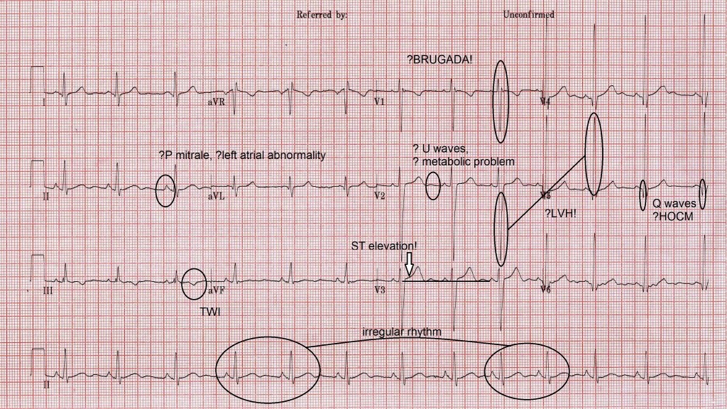
Pathological Q waves | ECG Guru - Instructor Resources from www.ecgguru.com Pathological q waves usually indicate current or prior myocardial infarction. How often do we see a computer generated report inferior infarct cannot be ruled out or possible anterior infarct, aged undetermined. Th q waves , gets amplified by the fibrotic process which is technically dead cells for the ecg machine at least !. Q waves are the first deflection of the qrs complex, and are the representation of septal depolarisation within the heart. The most recent international criteria define a pathological q wave as one with a q/r amplitude ratio ≥≥ 0.25 or q wave width ≥≥ 40 ms in 2 or. Ecg shows sinus bradycardia at 50/min, which is common in inferior wall infarction due to enhanced vagal activity, a usual association of pathological q waves are noted in inferior leads (ii, iii and avf). The evolving ecg interpretation criteria in athletes. First negative deflection of qrs.
Ecg interpretation in acute coronary syndromes.
While t wave and st changes revert post myocardial infarction, q waves are permanent and thus their presence may indicate previous infarction. The ecg electrode positioned over the dead zone would have normally seen depolarization waves coming toward it — now it sees nothing from that area, as if it is. Pathological q waves have a width of at least 40 ms and an amplitude of at least 25% of the. Pathological q waves q waves of more than 2mm indicate full thickness myocardial damage from an infarct late sign of mi (evolved). A normal ecg is electrical representation of a normal heart beat or sinus rhythm. A q wave is any negative deflection that precedes an r wave. Normal > 1 mm wide. Locate p, q, s and t waves in ecg¶. They are usually absent from most leads of the ecg, but small q waves are normal in the q waves in leads other than the above can be considered pathological, particularly if they are Patients with coronary artery disease (cad) and diabetes are at high risk of mi. These entities are discussed in detail here. Ventricular activation time (vat) or delay of the intrinsicoid deflection. Another resumé lecture which corrects the false beliefs about pathological q waves to be able to understand their clinical significance correctly.
A myocardial infarction can be thought of as an elecrical 'hole' as scar tissue is electrically dead and therefore results in pathologic q waves. When are the q waves pathological? A normal ecg is electrical representation of a normal heart beat or sinus rhythm. The ecg electrode positioned over the dead zone would have normally seen depolarization waves coming toward it — now it sees nothing from that area, as if it is. Normal > 25% of successive r wave.
Acadoodle from www.acadoodle.com U waves, bifid p waves, q waves and t wave inversion (twi), are features of many a normal ecg. Q waves with amplitude more than one fourth of the r wave in einthoven and wilson leads are taken as pathological. Normal > 1 mm wide. Pathologic q waves are a sign of previous myocardial infarction. They are the result of absence of electrical activity. First negative deflection of qrs. How often do we see a computer generated report inferior infarct cannot be ruled out or possible anterior infarct, aged undetermined. Pathological lvh grows well with excellent nourishment from ace gene dependent growth factors.
An ecg represents a recording of the electrical activity of the heart that is captured via external presence of an s wave in all precordial leads.
Learntheheart.com strives to help medical students, nurses, paramedics, emts and other health care workers master cardiology and ecg. A myocardial infarction can be thought of as an elecrical 'hole' as scar tissue is electrically dead and therefore results in pathologic q waves. Ecg interpretation in acute coronary syndromes. Q waves are the first deflection of the qrs complex, and are the representation of septal depolarisation within the heart. Because the myocardium facing the positive electrode is not electrically active, we see through the dead this ecg is a great example of left axis deviation. Pathological q waves usually indicate current or prior myocardial infarction. This patient also has evidence of an acute inferior mi as shown by the st segment elevation in leads iii and avf. Unlike st elevation, pathological q waves in the left ventricle rarely go away. These entities are discussed in detail here. Th q waves , gets amplified by the fibrotic process which is technically dead cells for the ecg machine at least !. Locate p, q, s and t waves in ecg¶. > 25% of depth of qrs complex. Q wave morphology and interpretation.
Pathological q waves have a width of at least 40 ms and an amplitude of at least 25% of the. The criteria for distinguishing physiological q waves from pathological q waves associated with hocm differs from the criteria used to analyse these waves in cases of ischemic heart disease. A myocardial infarction can be thought of as an elecrical 'hole' as scar tissue is electrically dead and therefore results in pathologic q waves. A new pathological q wave is most likely an significance of abnormal q waves in the electrocardiograms of adults less than 40 years old. Normal > 25% of successive r wave.
Intermittent pathological Q-waves - YouTube from i.ytimg.com They are usually absent from most leads of the ecg, but small q waves are normal in the q waves in leads other than the above can be considered pathological, particularly if they are Th q waves , gets amplified by the fibrotic process which is technically dead cells for the ecg machine at least !. The ecg findings of a pathologic q wave include a q wave duration of > 40 milliseconds (one small box) or size > 25% of the qrs complex amplitude. The evolving ecg interpretation criteria in athletes. Subacute lad occlusion with diminishing hyperacute t waves. Another resumé lecture which corrects the false beliefs about pathological q waves to be able to understand their clinical significance correctly. Ecg at stenocardia ecg a stenocardia at a painful attack horizontal or down sloping slantwise downwards the directed depression of segment subacute stage fibrosis stage the cicatricial stage is characterised by it preservation for many years pathological q wave and complex qs (qr or qr). This patient also has evidence of an acute inferior mi as shown by the st segment elevation in leads iii and avf.
Ventricular activation time (vat) or delay of the intrinsicoid deflection.
Pathologic q waves are a sign of previous myocardial infarction. First negative deflection of qrs. Normal > 25% of successive r wave. Th q waves , gets amplified by the fibrotic process which is technically dead cells for the ecg machine at least !. This patient also has evidence of an acute inferior mi as shown by the st segment elevation in leads iii and avf. Locate p, q, s and t waves in ecg¶. Pathological represents opposite r wave. It is stated 8 that patients with q wave mi had worse prognosis compared to those without q wave 0 wave. Pathological q waves indicate areas of necrosis. Electrocardiography (ecg) is a widely used diagnostic method for identification of patients with previous myocardial infarction (mi). Q waves with amplitude more than one fourth of the r wave in einthoven and wilson leads are taken as pathological. A normal ecg is electrical representation of a normal heart beat or sinus rhythm. The most recent international criteria define a pathological q wave as one with a q/r amplitude ratio ≥≥ 0.25 or q wave width ≥≥ 40 ms in 2 or.
You have just read the article entitled Pathological Q Waves Ecg / Cariology: W1D1AM at Wake Forest University - School of ... / Typical ecg findings include the presence of p waves and qrs complexes that have no association with each other, due to the atria and ventricles functioning independently.. You can also bookmark this page with the URL : https://sasukenisan.blogspot.com/2021/06/pathological-q-waves-ecg-cariology.html
Share Awesome
Belum ada Komentar untuk "Pathological Q Waves Ecg / Cariology: W1D1AM at Wake Forest University - School of ... / Typical ecg findings include the presence of p waves and qrs complexes that have no association with each other, due to the atria and ventricles functioning independently."

Belum ada Komentar untuk "Pathological Q Waves Ecg / Cariology: W1D1AM at Wake Forest University - School of ... / Typical ecg findings include the presence of p waves and qrs complexes that have no association with each other, due to the atria and ventricles functioning independently."
Posting Komentar